Image processing in the field of biology and medicine is crucial to a number of sectors. Research and industry, for example. More and more applications require image processing and analysis.
Several microscopy techniques are commonly used in public and private research. Each one generates customized analysis requirements, adapted to each project.
For example:
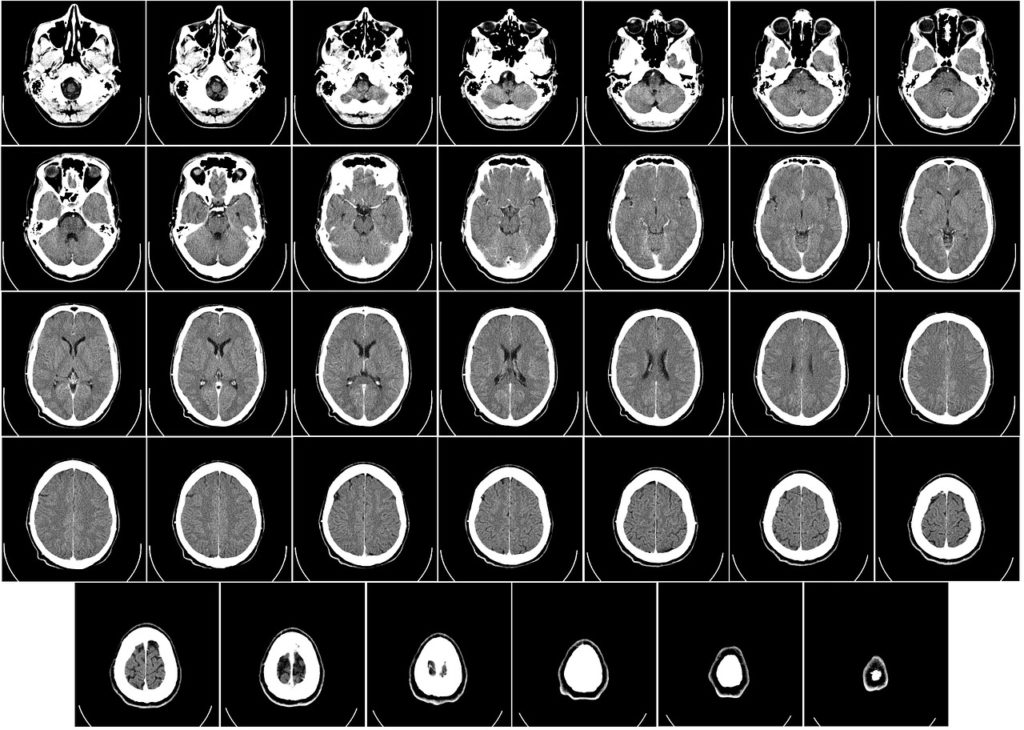
We also need to study images from other sources, such as MRI, PET, or simply cameras filming a patient, to gain access to information useful in research, diagnosis and treatment of disease.
Image analysis in Biology
Microscopic image analysis can be used to study a wide range of phenomena, such as :
- The distribution of certain molecules in tissues, as in spatial transcriptomics applications),
- Segmentation of certain objects at the level of single cells (such as organelles, DNA regions or condensates, which are useful for studying LLPS phenomena),
- Segmentation of organisms, such as bacteria,
- Cell classification (in a tissue or, for example, in a cell sorting application ),
- And the tracking of cells or other organisms.
Image enhancement in Biology
Often, complex imaging conditions mean that the images obtained must be prepared for use. Among the most common requirements are :
- Improved image quality,
- Correction of the offset between images taken within a time interval,
- Background noise reduction with denoising and deconvolution algorithms,
- Normalizing light intensity in the image,
- Chromatic aberration correction,
- ...
These improvements are now widely used in biology.
Medical decision support systems
The use of Deep Learning models in the medical field can aid decision-making and disease diagnosis, or help to improve and better protect patients. Several applications can be developed to this end.
Deep Learning for medical image enhancement
AI can help with decision-making at the time of diagnosis. Indeed, images from these diagnoses can be used to train AI models to recognize tumors. Current applications include improving the image quality of CT scans.
In this way, radiation doses can be reduced to minimize patient exposure. Signal loss is compensated for by algorithmic image enhancement.
A case in point: super-resolution
We can also cite an example of image enhancement with AI inference in the context of super-resolution.
The aim of super-resolution is to add information to a raw signal (in this case, pixels). In this way, we artificially upscale images to increase their resolution and thus facilitate medical analysis and interpretation.
For more information, we have already produced several articles on super-resolution:
In brief
As we have briefly described, image analysis is an indispensable tool in both public and private research, as well as in the medical field.
Our expertise in image processing, algorithm creation and the development of specific artificial intelligence models enables us to propose relevant solutions and support you at every stage of your project.
Finally, the fields of Biology and Medicine are very often linked to that of Fluid Mechanics. We are also developing particle-based optical diagnostics to characterize these flows.
Whether you're a research laboratory or a company, you can contact us for a solution tailored to your needs, particularly for image processing in the Biology and Medical fields.