What is flow cytometry?
Flow cytometry (FMC) is a reference technique for the analysis of particles suspended in a fluid. It enables the simultaneous measurement of several physical and biological characteristics of each particle. This is typically the case for cells or bacteria, at a flow rate of up to several thousand events per second.
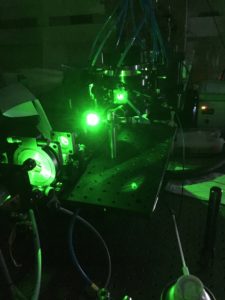
The principle is based on hydrodynamic injection of the sample into a stream. The aim is to force the particles to line up and pass through one or more focused laser beams. Each particle scatters light as it interacts with the beam. Specific fluorophores can mark it, enabling it to emit a fluorescent signal. Forward scattering (FSC) gives an estimate of cell size. Side scatter(SSC) provides information on internal complexity, such as cytoplasmic granularity. Fluorescence signals reflect the expression of specific biological targets.
These light signals are collected by a network of photodetectors (photodiodes, photomultipliers). They are then converted into electrical signals, digitized and analyzed in real time. Flow cytometry thus makes it possible to classify, quantify and sort cell subpopulations. This is done on the basis of several simultaneous criteria, with high sensitivity and reproducibility.
How does flow cytometry work?
A flow cytometer is structured around three main subsystems: fluidics, optics and detection electronics. These modules work in synergy to guarantee reliable, reproducible analysis of every event measured.
The fluidic system
The heart of the fluidic system is based on the principle of hydrodynamic focusing. The sample to be analyzed is injected into the center of a flow of physiologically neutral liquid. This forces the particles to flow in a single file. This configuration ensures that the events detected correspond to a single particle at a time. This avoids overlapping or counting errors.
The optical system
The aligned particles then pass into the analysis chamber. They are illuminated by one or more lasers of specific wavelengths. Incident light is either scattered or absorbed, then re-emitted as fluorescence if the particle is labelled. Collecting lenses and other optical elements direct the light to various detectors.
The two types of scattering, FSC and SSC, provide a physical estimate (size and complexity). Fluorescent signals, on the other hand, reveal biological or biochemical information about the particle, depending on the markers used.
The detection system
Photodiodes or photomultipliers (PMT) convert each optical signal into an electrical signal, then a system digitizes it. Downstream processing is carried out by specialized software that extracts the measured parameters.
Cytometer quality control includes regular calibrations using standardized fluorescent beads. This ensures stability and comparability of measurements over time.
Practical applications of flow cytometry
Flow cytometry is now indispensable in many areas of life sciences, thanks to its unique ability to rapidly analyze thousands of individual cells, with multiparametric resolution.
Immunophenotyping and hematology
One of the most widespread uses of CMF is the identification of cell subpopulations in blood or lymphoid tissue. Using fluorescently-labeled monoclonal antibodies, it is possible to precisely characterize lymphocyte types, hematopoietic stem cells or tumor cells. This approach is used in particular for diagnosing and monitoring leukemia, lymphoma and other hematological malignancies.
Cell cycle analysis and apoptosis
By marking DNA with intercalating dyes, CMF makes it possible to assess the distribution of cells in the different phases of the cycle, and to detect programmed cell death (apoptosis) through markers. Researchers routinely employ these analyses in oncology and pharmacology research, for example.
Cell sorting (FACS)
Some cytometers, known as sorters(Fluorescence Activated Cell Sorters), can physically separate cells of interest after their optical analysis, thanks to an electrostatic deflection system. This high-throughput sorting is essential for the purification of rare populations, such as stem cells or genetically modified cells, for downstream studies or therapeutic applications.
Applications in microbiology, bioprocessing and the environment
Researchers also use CMF to analyze bacterial populations, monitor viability in bioproduction cultures, detect pathogens or monitor the microbiological quality of water. Its speed and precision make it an invaluable tool for industrial laboratories and on-line processes.
Challenges and prospects
Although flow cytometry is a mature technology, it still faces a number of technical limitations, and is giving rise to numerous innovations designed to extend its analytical capabilities.
The main constraints include :
- Spectral compensation, necessary when several fluorochromes have overlapping emission spectra, which can lead to interpretation artifacts if incorrectly calibrated,
- Cellular autofluorescence, particularly in certain lines or primary samples, which can mask weak signals,
- Cell viability, often impacted by marking treatments or sorting conditions, which limits certain clinical or therapeutic applications,
- Data processing, which becomes more complex as the number of parameters measured increases, requiring advanced visualization and statistical analysis tools.
Recent advances aim to push back these limits:
- Spectral cytometry, which no longer uses fixed filters but analyzes the full spectrum of each fluorochrome, improves resolution and the ability to use many markers simultaneously,
- Imaging flowcytometry, combining flow and microscopy, enables advanced morphological analysis while maintaining high throughput,
- Integration with artificial intelligence, in particular machine learning, facilitates automatic recognition of cellular subpopulations in complex datasets,
- Lastly, microfluidic approaches are tending to miniaturize cytometers, making them compatible with portable analyses or integrated into on-chip diagnostic devices.
With these developments, flow cytometry is establishing itself as a pivotal technology in personalized medicine, cutting-edge biomedical research and industrial biotechnologies.
Would you like to find out more about our optical non-destructive diagnostics? Contact us to discuss your photonics needs.